STEREOTACTIC BREAST BIOPSY

A breast biopsy is the process of removing a small tissue sample from the breast for further evaluation in a laboratory. The doctors at Rose Imaging Specialists perform different types of breast biopsies, depending on the type and nature of the mass found within the breast.
Stereotactic Breast Biopsy is a type of core needle biopsy in which the radiologist uses a special computer to guide a needle to an abnormality seen on mammography. It employs a precise technique that uses digital imaging and is less invasive than a surgical biopsy.

WHY ARE STEREOTACTIC-GUIDED BREAST BIOPSIES NEEDED?

  • A mammogram shows a suspicious cluster of small calcium deposits.
  • The breast tissue is distorted and no corresponding finding is noted on ultrasound.
  • In the rare instance when a mass is seen on a mammogram, but is not able to be seen on an ultrasound.


BEFORE YOUR STEREOTACTIC BREAST BIOPSY

There are no diet restrictions prior to this procedure, but you may prefer a light meal so that you will be more comfortable.

In order to reduce the risk of bleeding during the procedure, we recommend that patients not take any aspirin or ibuprofen product (such as Advil or Motrin) for five days prior to the procedure. If you are on prescription blood thinning medication such as Coumadin, our staff can assist you in consulting your physician prior to scheduling this exam.

DURING YOUR STEREOTACTIC BREAST BIOPSY

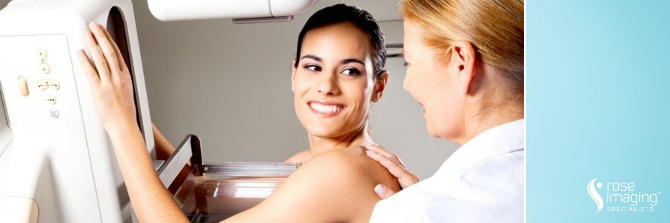
The first part of the procedure will seem much like your mammogram except that you will be sitting or lying down. Your breast will be compressed, usually somewhat less tightly than during a regular mammogram. An x-ray will be taken to confirm that the proper area of the breast is centered in the window in the compression paddle. When the position is ideal, more x-rays will be obtained. With the help of a computer, the exact positioning of the biopsy needle is determined from these images. Using this information, the radiologist will then position the device holding the biopsy needle.

Your breast will then be cleaned with antiseptic. Next, the radiologist will numb the part of the breast to be biopsied by injecting local anesthetic. This is done with a tiny needle, and you may feel a stinging at this point. After the local anesthetic has taken effect, the radiologist will make a skin nick through which the biopsy needle will be placed. Another pair of images will be taken to confirm the needle position. Once placement is confirmed, the tissue samples (cores) are acquired. Often the tissue samples are x-rayed to ensure they contain a representative sample of the area in question.

The entire procedure usually takes 30-60 minutes.