MRI-GUIDED BREAST BIOPSY

In MRI-guided breast biopsy, magnetic resonance imaging is used to help guide the radiologist’s instruments to the site of the abnormal growth for lesions only seen on MRI.

WHEN IS AN MRI-GUIDED BREAST BIOPSY PERFORMED?

An MRI-guided breast biopsy is most helpful when MR imaging shows a breast abnormality such as:

  • A suspicious mass not identified by other imaging techniques
  • An area of distortion
  • An area of abnormal tissue change

MRI guidance is used in three biopsy procedures:

Fine Needle Aspiration (FNA), which uses a very small needle to extract fluid or cells from the abnormal area.

Core Needle Biopsy (CNB) which uses a large hollow needle to remove one sample of breast tissue per insertion.

Wire Localization in which a guide wire is placed into the suspicious area to help the surgeon locate the lesion for surgical biopsy.

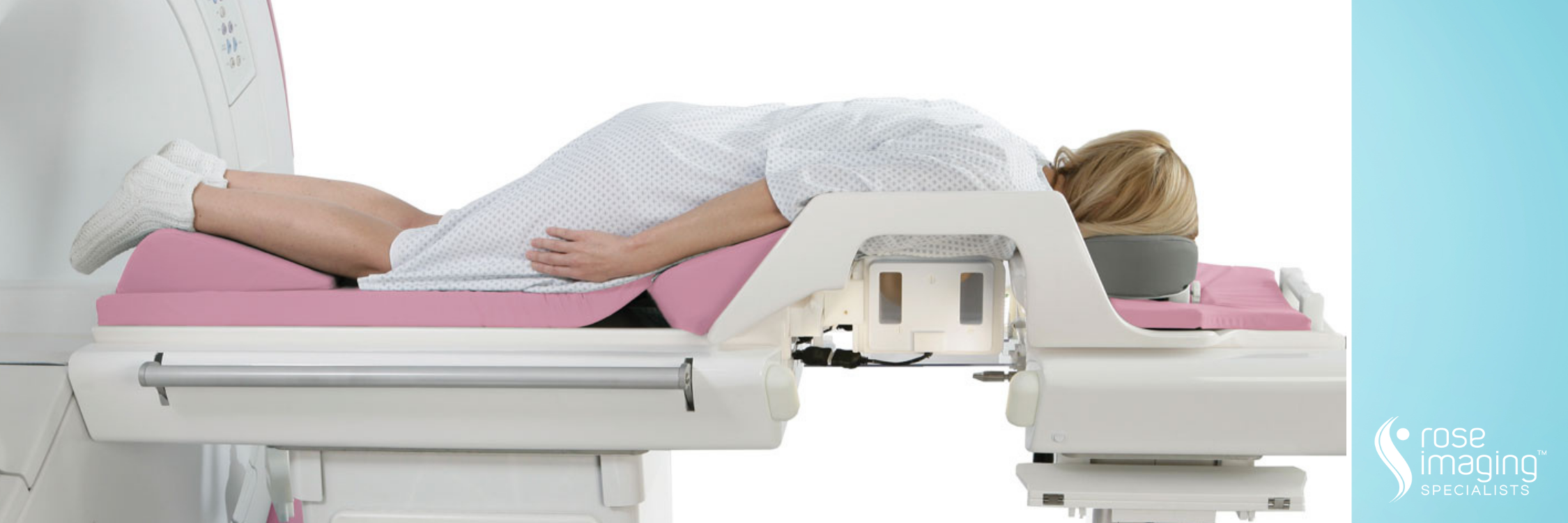
BEFORE YOUR MRI-GUIDED BREAST BIOPSY

Prior to a needle biopsy, you should report to your doctor all medications that you are taking, including herbal supplements, and if you have any allergies, especially to anesthesia. Your physician will advise you to stop taking aspirin or a blood thinner three days before your procedure.

Guidelines about eating and drinking before an MRI exam vary with the specific exam and also with the facility. For some types of exams, you will be asked to fast for 8-12 hours. Unless you are told otherwise, you may follow your regular daily routine and take medications as usual.

In order to reduce the risk of bleeding during the procedure, we recommend that patients not take any aspirin or ibuprofen product (such as Advil or Motrin) for five days prior to the procedure. If you are on prescription blood thinning medication such as Coumadin, our staff can assist you in consulting your physician prior to scheduling this exam.

The radiologist should also know if you have any serious health problems or if you have recently had surgery. Some conditions, such as severe kidney disease may prevent you from being given contrast material for an MRI. If there is a history of kidney disease, it may be necessary to perform a blood test to determine whether the kidneys are functioning adequately. You may want to have a relative or friend accompany you and drive you home afterward. This is recommended if you have been sedated.

Please notify our scheduling department and technologists if you believe you may be pregnant. MRI has been used for scanning patients since the 1980’s with no reports of any ill effects on pregnant women or their babies. However, because the baby will be in a strong magnetic field, pregnant women should not have this exam unless the potential benefit from the MRI is assumed to outweigh the potential risks.

DURING YOUR MRI-GUIDED BREAST BIOPSY

You will lie face down on a moveable exam table and the affected breast or breasts will be positioned into openings in the table. A nurse or technologist will insert an intravenous (IV) line into a vein in your hand or arm and the contrast material, gadolinium, will be given intravenously.

Your breast will be gently compressed between two compression plates, one of which is marked with a grid structure. Using computer software, the radiologist measures the position of the lesion with respect to the grid and calculates the position and depth of the needle placement.

The breast is cleaned and prepped.  A local anesthetic will be injected into the breast to numb it.  A very small nick is made in the skin at the site where the biopsy needle is to be inserted.

The radiologist then inserts the needle, advances it to the location of the abnormality and MR imaging is performed to verify its position. Depending on the type of MRI unit being used, you may remain in place or be moved out of the center of the MRI scanner.

Your MRI-guided biopsy usually takes 30 to 60 minutes.