BEFORE YOUR BREAST ULTRASOUND
- No fasting or sedation is required before the procedure.
- Dress in clothes that permit access to the area to be tested or that are easily removed.
A breast ultrasound uses high frequency sound waves to generate images of the tissues inside the breast. The sound waves pass through the breast and bounce back, or echo, from various tissues to form a picture of the internal structures. It is not invasive and involves no radiation or X-rays. The breast ultrasound can show all areas of the breast, including the area closest to the chest wall, which can be hard to study with a mammogram.
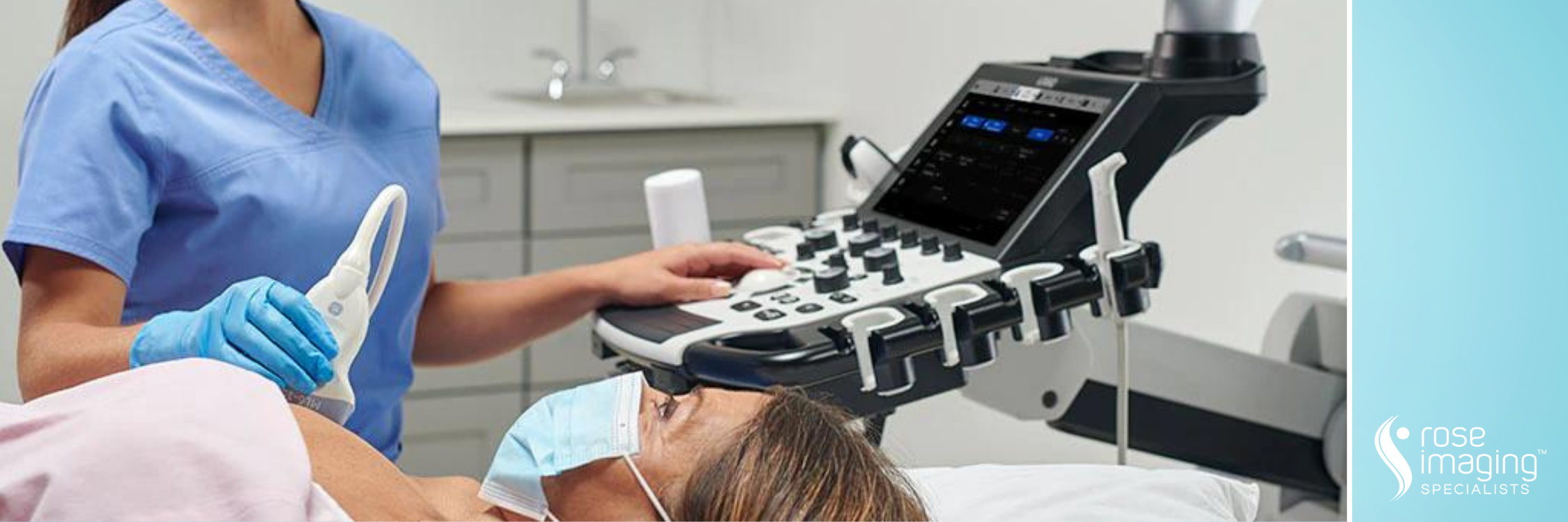
First, you will lie on your back with your arm raised above your head on the examining table. A clear water-based gel is applied to the breast to help the transducer make secure contact with the body and eliminate air pockets between the transducer and the skin. The sonographer (ultrasound technologist), followed by the radiologist, then presses the transducer firmly against the skin of the breast to obtain images for interpretation. The entire exam usually takes 20 minutes.
BEFORE YOUR BREAST ULTRASOUND
DURING YOUR BREAST ULTRASOUND