DUCTOGRAPHY

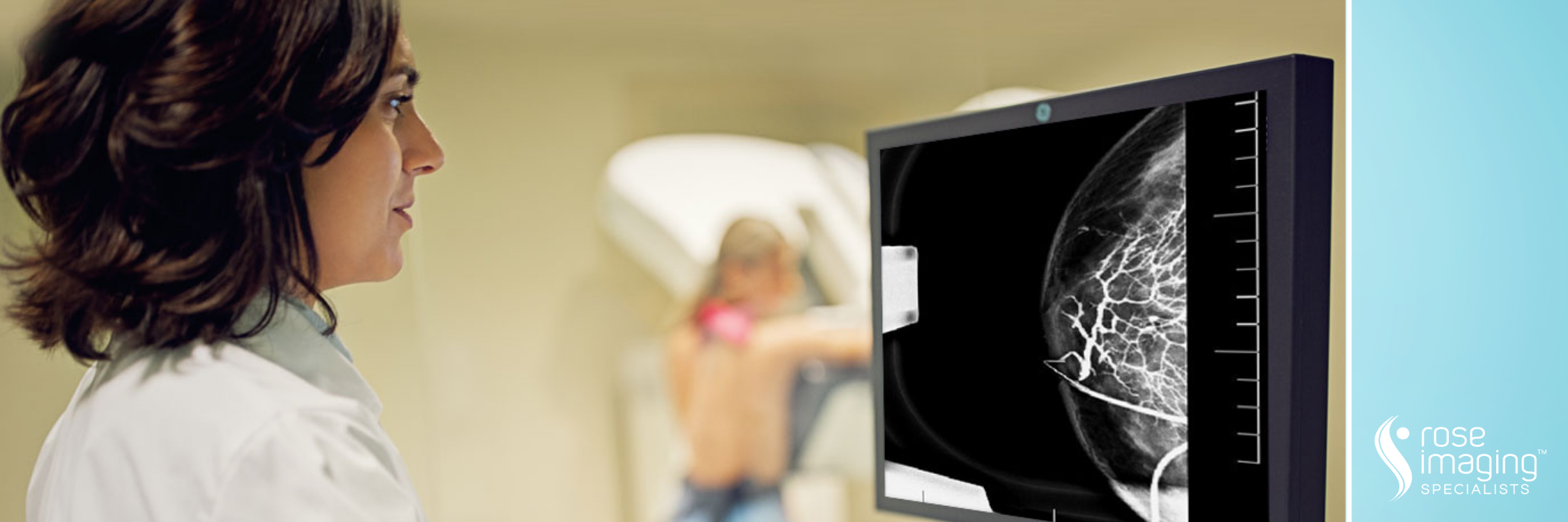
A ductogram, or galactogram, is a diagnostic exam used to identify and examine a duct—which is a small opening in the nipple—where nipple discharge has been observed. It is an x-ray examination that uses mammography, a low-dose x-ray system for examining breasts. These images are obtained by injecting contrast material into the discharging duct. These pictures are called galactograms and show the inside of the breast’s milk ducts.

The most common use of ductograms is to evaluate a woman who has a spontaneous bloody or clear discharge from her nipple and an otherwise normal mammogram and ultrasound.

Ductography is typically NOT called for in women with the following conditions:

  • A discharge that is milky, yellow, green, black or gray is usually not a cause for concern, especially if it comes from multiple ducts in the breast.
  • A discharge that is from both breasts in a woman who has not had children may indicate a side effect from a drug or may be related to a pituitary problem located in the brain.

BEFORE YOUR DUCTOGRAM

Very little preparation is necessary for this procedure. The only requirement is that the nipple not be squeezed prior to the exam, as sometimes there is only a small amount of fluid and it is necessary to see where that fluid is coming from to perform the exam.

You should inform your physician of any medications you are taking and if you have any allergies, especially to barium or iodinated contrast materials. Also inform your doctor about recent illnesses or other medical conditions. As in mammography, do not wear deodorant, talcum powder or lotion under your arms or on your breasts on the day of the exam. The residue left on your skin by these substances may interfere with the X-rays.

DURING YOUR DUCTOGRAM

You will be asked to undress above the waist and lie on your back with your arms above your head. Topical anesthetic cream will be placed on the nipple to numb the area. The radiologist will identify the duct using a magnifying glass and will insert a tiny catheter into the duct. The duct is a natural opening that should be dilated due to the discharge.

Once the catheter is inserted, the radiologist will fill the duct with a radio-opaque dye that shows up on a mammogram. A mammogram is done after the injection of dye to show the outline of the duct. Once the catheter is removed, the duct will discharge the dye.

The entire exam usually takes 30 minutes.